Breast Biopsy
What is a breast biopsy?
A biopsy is a small piece of tissue that is removed and checked in a lab. For a breast biopsy, breast tissue may be removed with a special biopsy needle. Or it may be removed during surgery. It is checked to see if cancer or other abnormal cells are present.
Why might I need a breast biopsy?
 |
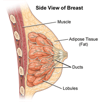
| Click Image to Enlarge |
Breast biopsies may be done:
-
To check a lump or mass that can be felt (is palpable) in the breast
-
To check a problem seen on a mammogram, such as small calcium deposits in breast tissue (microcalcifications) or a fluid-filled mass (cyst)
-
To evaluate nipple problems, such as a bloody discharge from the nipple
-
To find out if a breast lump or mass is cancer (malignant) or not cancer (benign)
A lump or other area of concern in the breast may be caused by cancer. Or it may be caused by another less serious problem.
There may be other reasons for your doctor to recommend a breast biopsy.
Types of breast biopsies
There are several types of breast biopsy procedures. The type of biopsy that you have will depend on the location and size of the breast lump or area of concern.
Biopsies may be done under local or general anesthesia. For local anesthesia, medicine is injected to numb your breast. You will be awake, but feel no pain. For general anesthesia, you will be given medicine to put you into a deep sleep during the biopsy.
Types of breast biopsies include:
-
Fine needle aspiration (FNA) biopsy. A very thin needle is placed into the lump or area of concern. A small sample of fluid or tissue is removed. No cut (incision) is needed. An FNA biopsy may be done to help see if the area is a fluid-filled sac (cyst) or a solid lump.
-
Core needle biopsy. A large needle is guided into the lump or area of concern. Small cylinders of tissue, called cores, are removed. No cut is needed.
-
Open (surgical) biopsy. A cut is made in the breast. The surgeon removes part or all of the lump or area of concern. In some cases the lump may be small, deep, and hard to find. Then a method called wire localization may also be used. For this, a thin needle with a very thin wire is put into the breast. X-ray images help guide it to the lump. The surgeon then follows this wire to find the lump.
Special tools and methods may be used to guide the needles and help with biopsy procedures. These include:
-
Stereotactic biopsy. With this method, a 3D image of the breast is made using a computer and mammogram results. The 3D image then guides the biopsy needle to the exact site of the breast lump or area of concern.
-
Vacuum-assisted core biopsy. A small cut is made in the breast. A hollow tube or probe is inserted through the cut. It is guided to the breast lump or mass by MRI, X-rays, or ultrasound. The breast tissue is gently pulled into the probe. A spinning knife inside the tube cuts the tissue from the breast. Several tissue samples can be taken at one time.
-
Ultrasound-guided biopsy. This method uses ultrasound images of the breast lump or mass. These images help guide the needle to the exact biopsy site.
What are the risks of a breast biopsy?
All procedures have some risk. Some possible complications of a breast biopsy include:
-
Bruising and mild pain at the biopsy site
-
Prolonged bleeding from the biopsy site
-
Infection near the biopsy site
If the biopsy is done using an X-ray, the amount of radiation used is small. The risk for radiation exposure is very low.
You may have other risks depending on your specific medical condition. Be sure to discuss any concerns with your healthcare provider before the biopsy is done.
How do I get ready for a breast biopsy?
Your provider will explain the procedure to you. Ask any questions you have about the procedure.
-
You will be asked to sign a consent form that gives your permission to do the procedure. Read the form carefully and ask questions if something is not clear.
-
Generally, you don’t need to prepare for a biopsy if your breast is numbed (local anesthesia) but you are awake. But if you are given medicine to put you into a deep sleep (general anesthesia), you must not eat or drink anything (fast) for some time before the surgery. Your surgeon will give you specific instructions.
-
Don’t use lotion, cream, powder, deodorant, or perfume on your arm, underarm, or breast on the day of the procedure.
-
Tell your provider if you are pregnant or think you may be pregnant.
-
Tell your provider if you are sensitive to or allergic to any medicines, latex, tape, and anesthesia medicines (local and general).
-
Tell your provider about all the medicines you take. This includes both over-the-counter and prescription medicines. It also includes vitamins, herbs, and other supplements.
-
Tell your provider if you have a history of bleeding disorders. Let your provider know if you are taking any blood-thinning medicines, aspirin, ibuprofen, or other medicines that affect blood clotting. You may need to stop taking these medicines before the biopsy.
-
If you have a sedative or general anesthesia, make sure you have someone drive you home afterward. You will not be able to drive after the biopsy.
-
Your healthcare provider may have other instructions for you based on your medical condition.
What happens during a breast biopsy?
A breast biopsy may be done in a provider’s office. It may be done on an outpatient basis, which means you go home the same day. Or it may be done as a hospital stay. Some types of biopsies only require local anesthesia. For other types, general anesthesia is needed. Procedures may vary depending on your condition and your provider’s practices.
Generally, a needle breast biopsy follows this process:
-
You will be asked to remove any clothing from the waist up, and given a gown to wear.
-
You will lie down or sit up. This depends on your provider’s preference and if X-ray or ultrasound guidance will be used.
-
The skin over the biopsy site will be cleaned with a sterile solution.
-
When a local anesthetic is used, you will feel a needle stick when the medicine is injected. You may feel a brief stinging feeling. The biopsy will not start until the area is numb.
-
When ultrasound is used, the probe will be placed on your breast to find the breast lump or mass.
-
When stereotactic imaging is used, you will lie face down with your breast placed in an opening on the table. A computer will find the exact site of the breast lump or area of concern.
-
You will need to lie still during the procedure. But you won’t need to hold your breath.
-
The provider will place the needle right into the biopsy site or through a tiny cut (incision). He or she will remove a sample of tissue or fluid. You may feel pressure when the doctor takes the sample.
-
Pressure will be applied to the biopsy site until the bleeding stops.
-
If needed, the opening will be closed with stitches or adhesive strips.
-
A sterile bandage or dressing will be applied.
-
The tissues will be sent to the lab for examination.
Generally, an open breast biopsy will follow this process:
-
You will be asked to remove clothing and given a gown to wear.
-
An IV (intravenous) line may be started in your arm or hand.
-
You will be placed on the operating table.
-
You may be given medicine (a sedative) to relax you.
-
If local anesthesia is given, you will feel a needle stick to numb your breast tissue.
-
If general anesthesia is given, the anesthesiologist will keep checking your heart rate, blood pressure, breathing, and blood oxygen level during the surgery.
-
In some cases, a special wire will be placed into the lump before the biopsy, guided by X-ray images. This is to help the doctor find the breast lump or mass. Other X-ray-guided methods may be used.
-
The skin over the surgical site will be cleaned with a sterile solution.
-
A small cut will be made in your skin and underlying breast tissue until the lump or mass is exposed.
-
A part of the lump or the full lump will be removed.
-
The opening will be closed with stitches or adhesive strips.
-
A sterile dressing or bandage will be applied.
-
The tissue will be sent to the lab for exam.
What happens after a breast biopsy?
How long it takes to recover will depend on the type of biopsy you had and the type of anesthesia used.
If you had general anesthesia, you will be taken to the recovery room, where you will be watched closely. Once your blood pressure, pulse, and breathing are stable and you are alert, you will be taken to your hospital room. Or you will go home. If the biopsy was done on an outpatient basis, you should plan to have another person drive you home.
If you had local anesthesia, you will be able to go home after you have completed the recovery period.
At home
Keep the biopsy area clean and dry. If stitches were used, they will be removed during a follow-up office visit. If there are no stitches, you may remove the bandage or dressing when instructed to do so, and bathe as normal.
The biopsy site may be sore for several days after surgery. Take a pain reliever as recommended by your doctor. Aspirin or certain other pain medicines may increase your chance of bleeding. Be sure to take only recommended medicines. Wearing a supportive bra may help with mild pain.
You may go back to your normal diet unless your doctor advises you differently. You may be told to avoid strenuous physical activity for a few days.
Call your healthcare provider if you have any of the following:
-
Fever or chills
-
Redness, swelling, bleeding, or other drainage from the biopsy site
-
Increased pain around the biopsy site
Your healthcare provider may give you other instructions, depending on your situation.
Next steps
Before you agree to a test or procedure, make sure you know:
-
The name of the test or procedure
-
The reason you are having the test or procedure
-
What results to expect and what they mean
-
The risks and benefits of the test or procedure
-
What the possible side effects or complications are
-
When and where you are to have the test or procedure
-
Who will do the test or procedure and what that person’s qualifications are
-
What would happen if you did not have the test or procedure
-
Any alternative tests or procedures to think about
-
When and how will you get the results
-
Who to call after the test or procedure if you have questions or problems
-
How much you will have to pay for the test or procedure